Images in Early Modern Scientific Books
Interest in the role of printed images within the discipline of history of science can be traced back to the paper by Martin Rudwick (1976), in which he showed how a visual language for the newly emerging discipline of geology in the early nineteenth century was forged from pre-existing forms of illustration techniques and conventions, within the economic and technical constraints of producing printed images of the time.1 Historians of science have regularly and profitably integrated scholarly methods and insights from the history of the book, and printed images in particular have been included in more recent analyses of specific features of images in the service of scientific knowledge, or “epistemic images”.2 My aim in this paper is to offer a brief and necessarily selective survey of the state of scholarship on images in early modern scientific books, with a focus on the image’s relationship to the text as well as to the object. It would be foolhardy to attempt any grand syntheses, given that different styles and functions of images existed in the early modern period according to different disciplines and genres.3 Nevertheless, in certain cases, it is possible to show that attention to an aspect dear to Henri-Jean Martin, “mise en page” – how text and image worked together on a page and within a physical book – helps to understand how scientific objects and arguments were formed on the page in the early modern period.4
It is helpful to recall that naturalistic techniques, however convincing they may appear to the eye, do not in themselves guarantee that an object depicted by such techniques actually existed or that direct observation of that object took place.5 If a naturalistic style of representation should be best understood as “rhetoric of the real”, as suggested by Kemp, how do we deal with those depicted objects? Does it mean that independent, scientific corroboration is required of objects depicted in early modern scientific books? Such corroboration of objects depicted in the past with modern equivalents has at times been helpful to the historian, paradoxically when discrepancies have been detected. Thus, Andreas Vesalius’s inclusion of very uncommon bone structures in his De humanis corporis fabrica has highlighted his commitment to teleological reasoning.6 Conrad Gessner’s image of a non-existent toucan in his Historia aimalium draws attention to his method of compilation using objects as well as textual description.7 The figures of the surface of the Moon in Galileo Galilei’s Sidereus Nuncius do not match exactly the actual views of the Moon at the corresponding phases because he had merged his drawings in order to fit them into the limited space available in the tract he was in a rush to print.8 In fact, a crater of an exaggerated size in the image helped his argument that the surface of the Moon was rugged, not smooth, based on an analogy with the valley of Bohemia. Rather than dismissing these discrepancies between a modern object and a past image as inaccuracies of scientific knowledge of the past, they can be treated as opportunities to examine more closely past functions of these images, often articulated in the text that the images accompanied.
One of the more obvious functions of printed images for the study of nature was to fix and preserve ephemeral things on a page. For example, features of plants that grow only during a particular time of the year or under a particular climate could be captured on a page for readers to examine at their leisure, at any time or place. Such a possibility was realized when a visually enterprising printer, Johannes Schott, commissioned artists (including Hans Weiditz) to draw images of plants around Strasbourg, and asked Otto Brunfels to write a text for those images.9 The title of the publication, Herbarum vivae eicones (1530) rightly emphasized its lively images as the distinguishing feature of the book. Brunfels’s text sought to match the depicted plants with medicinal plants mentioned by ancient authors. The fact that a plant for which a classical equivalent could not be found was called “nuda herba”, despite its vernacular name being known, reflects Brunfels’s interest in matching classical “words and things”.10 Earlier humanist scholars had sought to identify contemporary equivalents of classical plants by examining philology, manuscript transmission as well as morphological similarities with known plants, but they had not used images when discussing the identity of classical plants.11 The images in Vivae eicones herbarum introduced a visual dimension to this humanist study of plants by picturing objects for which an appropriate name had to be found. While Brunfels’s task was to find classical names for the plants found in the environs of Strasbourg, William Turner, an English humanist physician, was looking for English names in order to introduce classical medical botany into England. He had seen the woodcut of the narcissus in Brunfels’s Eicones, but could not find its English name until one day he saw a little girl carrying a bunch of flowers. “Those are Narcissi”, he thought, and asked the girl for its name, but she did not know. Nor did those living nearby know its name. They suggested “laus tibi”, which Turner found out soon afterwards was used for asphodel, not for narcissus. He showed the flowers he had obtained from the girl to an old man who was knowledgeable about plants, who said that it was a “French Gillyflower”.12 Turner had made a mental, visual match between the woodcut of Vivae eicones herbarum and the girl’s flower, which eventually helped him find the English name of a classical plant identified by Brunfels. An image thus helped to bridge plants and names in an environment away from the original locale, and also enabled one reader to build on another scholar’s identification.
The Latin title of Brunfels’s work, Herbarum vivae eicones ad naturae imitationem, summa cum diligentia et artificio effigiatae, indicated that the images were made with great diligence and skill “in imitation of nature”. As is well known, “imitation” in this period could take many forms, from the copying of every detail of how a particular thing is found to be, to a representation that corrects and perfects imperfections and individual idiosyncrasies.13 The attention to detail of the particularities of a singular object was a mode of representation that was often identified as “imago contrafacta”, a popular form of representing natural anomalies and abnormalities on single-sheet broadsides.14 The woodcuts of Strasbourgois plants in Herbarum vivae eicones are in this “contrafactum” style of imitating nature, as it recorded bent stems and leaves with holes and tears. This should be distinguished from another phrase used in the period, “ad vivum”, which first appeared in a title of a printed book to describe Hans Holbein the younger’s illustrations for the Old Testament, Historiarum veteris instrumenti icones ad vivum expressae (1538). Here “ad vivum” must have just meant vivid images that would impress the viewers, rather than images made “from the life”. As recent scholarship has shown, “ad vivum” in this period had multiple meanings and connotations, and did not necessarily mean that an image was made from direct observation.15 This is consistent with the fact that there was not yet any consensus in the first half of the sixteenth century as to what was involved in a “scientific” form of looking at an object directly and attentively – “observation” certainly did not carry the modern connotation of scientific observation.16 Nor was the word “autopsia” (seeing for oneself) deployed in describing how scientific images had been made, possibly because of its negative association with the Empirical sect criticised by Galen.17 Thus, images in early modern scientific publications were called “figura”, “picture”, “effigies”, or “icons”, words that were commonly used to describe all manner of images.18
Leonhard Fuchs also styled his images as “ad naturae imitationem” on the title-page of his De historia stirpium (1542). As several scholars have noted, the style of the woodcuts in his book pushed in the opposite direction from Brunfels’s “contrafactum” images, into an idealised form that Fuchs described as “absolutissima”.19 The plants depicted in his book show no blemishes, included all stages and parts of a plant, and when the images were coloured, they also showed subspecies. They are not portraits of individual specimens observed by an artist at a particular time or place. In fact, it is not possible to come across a plant exhibiting several subspecies in one bush or all its stages of growth at a single moment. In other words, Fuchs’s images are universalized objects that do not exist in nature as such. To the extent that “scientia” of the period dealt with generalized, rather than particular objects at the time, it meant that it was on the page that the object of scientific investigation existed.20
Fuchs’s project was similar to Brunfels’s in that it was about identifying classical medicinal plants in contemporary plants. Fuchs emphasized that his identification was based on morphological correspondence, and thus each plant was provided with a different woodcut in his book. This was a direct challenge to contemporary practices of printers who re-used woodcuts to illustrate different objects or different books. Repeated use of the same woodcut for different plants was, according to Fuchs, a sign of a printer’s greed that jeopardized medical knowledge.21 Not everybody had the financial resources as Fuchs did to create afresh so many woodcuts, and object-specific images did not necessarily become the norm in “scientific” publications in this period.
Fuchs had written his text with a coloured woodcut in mind.22 Conrad Gessner also expected coloured woodcuts in his Historia animalium (1551-58) and De rerum fossilium, lapidum et gemmarum… figuris (1565). By exploiting the potential of the printed book to be coloured, authors could extend the effectiveness of their images to convey more information about variability of species, but they were also limiting the effectiveness of their statements, given that not all copies of their book were sold coloured. Copies coloured after an exemplar could be supplied by printers in some cases, as was the case with Gessner’s Historia animalium, but they cost two to four times the price of an uncoloured copy.23 When the colouring was left in the hands of the owners, a bewildering variety of colouring schemes ensued, as was the case with the copies of Pierre Belon’s Histoire de la nature des oyseaux (1555).24 Colouring of images thus remained a challenge to control or standardise in the sixteenth century.
Few authors in the sixteenth century succeeded in exerting control over their books as Andreas Vesalius did.25 The images in Vesalius’ Fabrica are well known for being well crafted and staged artistically.26 They are not documentary records of bodies of individuals dissected in the dissection hall. Instead, they show the canonical body of a “perfect man (homo absolutus)” without individual variations or pathological anomalies.27 Vesalius supervised closely the artists who made the images to ensure that they reflected the details he drew attention to in his textual description.28 Full-length figures of anatomised bodies occupying an entire page were keyed with letters, followed by names and descriptions of the parts under each letter. Smaller figures were placed within chapters that described how those parts were related and functioned, and even smaller figures and diagrams were placed in the outer margins to illustrate a particular point in the text. These well-known ways to link images and text were used by Vesalius to good effect, in order to present his view of the human body. The reader was informed through the text that Vesalius had deliberately included a muscle from a dog on a human figure or an erroneous picture of the heart, so that he could show Galen’s mistakes. Nor was it possible to understand the textual explanations without reference to the images. Vesalius was perhaps unusual for his time in exploiting the internal margins of the page, which were used to refer exclusively to the images (the outer margin was also used, in the traditional way of summarizing the main points of each section). A superscript alphabet letter was keyed in the text, and under that letter in the internal margin, locations of images of the relevant anatomical structure were listed. This guided readers to leave the page of the text, and look over to the figures elsewhere in the book. A cue to look up an image could be very frequent – as much as twenty to thirty times a page. This system meant that Fabrica was not a book to be read from the beginning to the end in one direction. The pages were to be turned backwards or forwards, to ensure that the text and image were integrated in the mind of the reader, to form knowledge about the human body. The fact that the image and the text were meant to work together in a book to produce “scientific” knowledge about an object implies that images in some early modern scientific books were not meant to be “books of the unlearned”. Rather, these images presupposed proficiency in at least Latin, and preferably also in Greek.
At least one reader responded to the close connection between text and image woven by Vesalius [ill. 1]. The Regius professor of medicine at Cambridge, Thomas Lorkyn, for example, added into his copy of the second edition of Fabrica (1555) some page numbers to parts of the body in the image so that they pointed to the location of the textual description of the relevant anatomical structure.29 While the internal margins of a text pointed the reader to the corresponding images, it was not possible to get to the relevant text from the images, so Lorkyn had made his own index to the text on the images.
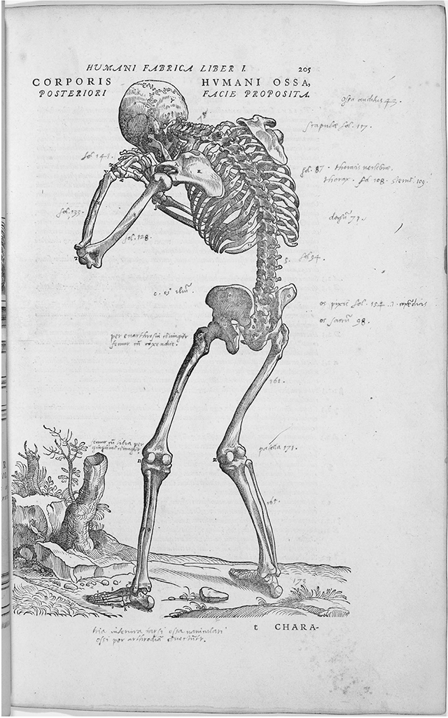
Ill. 1. The Regius professor of medicine at Cambridge, Thomas Lorkyn (1528-1591), annotated this image in his copy of Andreas Vesalius’s Fabrica (1555) as an index to the textual descriptions of the corresponding parts of the skeleton (Cambridge University Library, N*.1.1(A)).
Vesalius’s images were woodcuts, which were readily integrated with the text on the page. When Felix Platter printed his De corporis humani structura et usu (1583), copying Vesalius’s figures in intaglio, he decided to divide the text and image into separate volumes. This would have meant less frequent turning of the pages, and the ability to keep the text and image side by side. Intaglio could achieve finer lines in a smaller space than woodcut, but integrating intaglio images with the text on the page required putting the sheet of paper through two different presses. This was how diagrams in Galileo’s Saggiatore (1623) were set. In contrast, in Orazio Grassi’s reply to Galileo, Ratio ponderum librae et simbellae (1626), all the diagrams needed in Grazzi’s argument were engraved on a single plate [ill. 2], which was of course more economical. Interestingly, at the bottom of the sheet, it was written: “This little page of figures, in order that it may be used profitably, should be attached to the edge of a page at the beginning of this volume so that when folded out, it projects outside [of the book], as is shown here in the twelfth figure, and when folded up, it may be put away.” Figure 12 is a reflexive image of the book of which it is a part, and indicates how a sheet can be left folded out while the reader read the text. The fact that this convention is written out suggests that it was perceived to be a somewhat unusual arrangement at the time. It certainly suggests some concern to keep the text and image closer together. It was a convention that was picked up in the Royal Society’s Philosophical Transactions later in the seventeenth century. These different ways of keeping the text and image together highlight the assumption that the close working of the two were important in scientific books.
An image as a composite scientific object, and its interdependence with the text are features that also characterized scientific books in the later seventeenth century, in Robert Hooke’s Micrographia, for example. The book was intended from the beginning to be dedicated to Charles II by the Royal Society, and Robert Hooke submitted drawings regularly in 1663 to the meetings of the Royal Society where they were approved by the fellows attending the meetings.30 The images in Micrographia, though many of them set in a circle to simulate the field of vision of a microscope, were not snapshot views from a single observation – three-dimensional structures could only be determined by adjusting the focus of the microscope several times.31 Furthermore, as Hooke explained in the preface, the object needed to be viewed several times and under varying lighting conditions before he could make a drawing. Hooke’s images were therefore a composite of multiple observations. Microscopic images by definition also introduced the problem of scale; while nobody needed a scale on a page for Vesalius’s human body, a microscopic view of natural objects needed one. Hooke introduced scale bars in his images, but perhaps more effective to the reader was showing figuratively the actual size of an object right next the enlarged microscopic image, to show what an intricate pattern could be found in such a small structure [ill. 3].
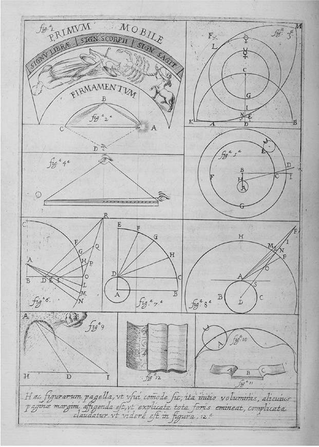
Ill. 2. Orazio Grassi, Ratio ponderum librae et simbellae (1627), sheet of diagrams. Fig. 12, the middle figure in the bottom row shows how the sheet should be glued to the edge of a page so that it can be consulted while the book is open (University of Oklahoma Libraries, History of Science Collections).
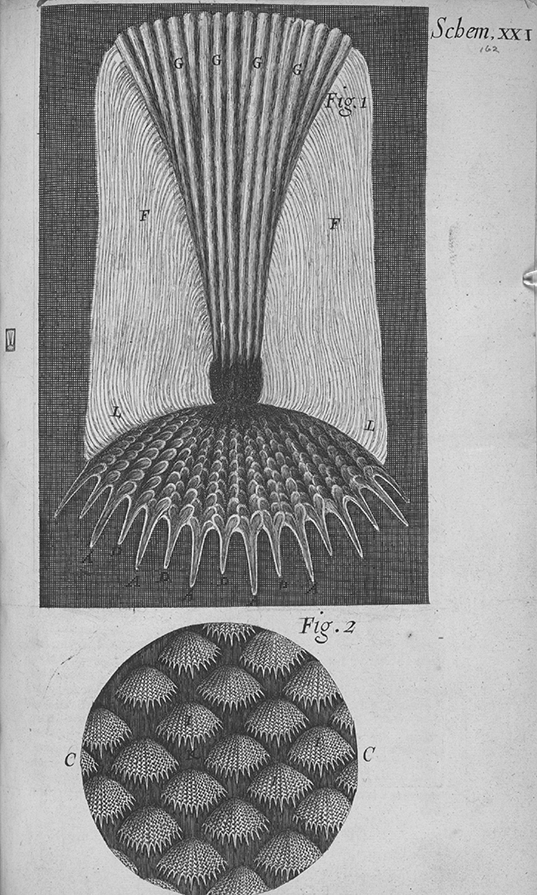
Ill. 3. Robert Hooke, Micrographia (1665), scheme XXI, etched and engraved, the fish scale in the left-hand margin is 7 mm high. Its enlarged image, labeled “Fig. 1”, is 18.9 mm high. (British Library, 435.e.19).
Though Micrographia was not the first printed book on microscopic observations and microscopes had been in circulation from about the 1620s, Hooke still faced a challenge that microscopic views of familiar natural objects were still unfamiliar to a general audience.32 He began by showing that human-made things that appeared to the naked eye to have perfect shapes, such as a circular full stop or a sharp razor edge, were in fact imperfectly shaped when viewed under the microscope. In contrast, nature’s various objects displayed under the microscope unexpected patterns and regularity. These microscopic structures of natural objects were so unfamiliar that they were impossible to be identified without the text, which also explained how the minute structures contributed to texture, springiness or other physical features of the object. The text made references back to the intaglio images that had been printed on separate sheets of paper and inserted near one of the references to the images, but because references to images were extensive in the text, it meant that the reader had to locate the relevant sheet. Most of the plates in Micrographia were much larger than the height of the page (c. 28 cm) of the book, and were kept folded in the book. When readers wanted to turn to the images in the book, they thus had to unfold manually a sheet of paper – to reveal in one case an image of a louse almost half a meter long. This was most probably a deliberate design decision, to surprise and impress the reader of visions of a microscopic world. At least one reader was impressed, as Christiaan Huygens exclaimed that the flea was the size of a small cat.33
One contemporary reader who understood that Hooke’s argument consisted of both image and text was Isaac Newton [ill. 4]. When he read the Micrographia, he took notes on the text and copied out the images. Historians of science have been examining closely the importance of note-taking and reading practices, and the extent to which it is possible identify “scientific” forms of such practices.34 It is very likely that Newton was in the minority of readers of Micrographia in copying out images alongside text. It does suggest, however that some scientific authors and readers of early modern scientific books understood that image and text worked together. By taking seriously what the pages in a scientific book look like, how they functioned within a physical book, and how they were read, it is possible to appreciate how in some scientific books text and image worked together to create objects of scientific knowledge, and shape scientific claims and arguments in the early modern period.
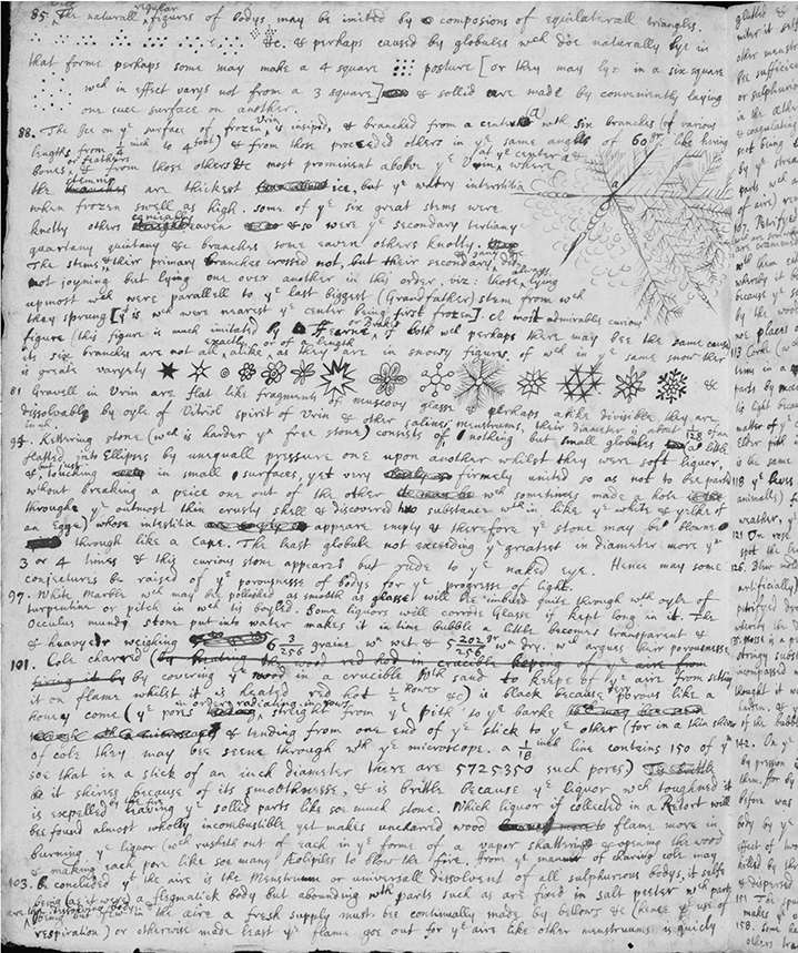
Ill. 4. Isaac Newton’s notes on Hooke’s Micrographia (Cambridge University Library, Add MS 3958, 2v).
Not all scientific books carried illustrations. Not every scientific author could exercise control over mise en page in the way that Vesalius, Fuchs or Hooke did. Not every scientific image remained flat on the printed page. Some were meant to be cut-up and glued together, to simulate anatomised bodies, planetary motions, or sun-dials.35 Most importantly, images quickly acquired a life of their own.36 The practice of copying and recopying images was pervasive among early modern printers, which meant that printed images were swiftly decoupled from the original text, and connected to another or no text at all.37 Copying a pre-existing image was a cost-cutting exercise on the part of printers, but in England, it appears to have been more financially viable to rent woodblocks from the Continent, possibly because there was a shortage of skilled woodcutters. For William Turner’s A new herball (1551), the printer Steven Mierdman rented one third of woodblocks from the Birckman firm that had been created for an octavo edition of Fuchs’s De historia stirpium (Paris, 1549). For John Gerard’s Herbal (1597), the printer John Norton used more than two thousand woodblocks originally cut for a pictorial album of plants by Jacobus Theodorus Tabernaemontanus (1588-1591) by the Frankfurt printer Nicolas Bassaeus. The second edition of Gerard’s The Herball, revised by Thomas Johnson (1633) was illustrated with 2791 woodblocks that the bookseller Richard Whitacker borrowed from the Plantin-Moretus firm in Antwerp.38 As it turns out, the woodcuts by Bassaeus used for the first edition of Gerard’s Herbal were copies of another album of plants published by Plantin nine years earlier. This meant that the first and the second editions of Gerard’s herbal had inverted woodcuts, the one in the first edition being a copy of those in the second edition.
Images in scientific books in the early modern period are ultimately part of a wider visual culture of science. Recent scholarship has also reminded us that they are only the tip of the iceberg of images of nature that were made and circulated in the early modern period.39 There are numerous sketches and watercolours of naturalia that belonged to various scholars and collectors of the period. Some of them were the original drawings from which printed images were made, others were copies or adaptations of printed images, and yet more could be identified as having been made after live samples, dried specimens or stuffed examples. The finely finished state of many suggest that they were not sketches done “in the field”, but an image developed from such sketches, and something artists spent time to refine and finish for collectors.40 Further questions remain, such as the exact form of collaboration between authors and graphic craftsmen; the relationship between printed images and observational practices as they became increasingly complex; whether it is possible to identify the emergence of a “scientific” visual culture, and the role of print in it.41
____________
1 Martin Rudwick, “The emergence of a visual language for geological science 1740-1840”, History of Science, 14, 1976, p. 149-195; for its delayed reception among historians of science, see Sachiko Kusukawa, “Classics from this journal: Martin Rudwick’s ‘The emergence of a visual language for geological science 1760-1840’”, History of Science, 54, 2016, p. 98-104.
2 For history of science and history of the book, see for example, Adrian Johns, The nature of the book, Chicago, Chicago University Press, 1999; Books and the sciences in history, ed. Marina Frasca Spada, Nicholas Jardine, and Silvia De Renzi, Cambridge, CUP, 2000; For an analysis of images in history of early modern science, see Non-verbal communication in science prior to 1900, ed. Renato G. Mazzolini, Firenze, Olschki, 1993; Picturing knowledge: Historical and philosophical problems concerning the use of art in science, ed. Brian S. Baigrie, Toronto, University of Toronto Press, 1996; The power of images in early modern science, ed. Wolfgang Lefèvre, Jürgen Renn, and Urs Schoepflin, Basel, Birkhauser, 2003; Picturing machines 1400-1700, ed. Wolfgang Lefèvre, Cambridge, MA MIT Press, 2004; Christoph Lüthy and Alexis Smets, “Words, lines, diagrams, images: Towards a history of scientific imager”, Early Science and Medicine, 14, 2009, p. 398-439; Art and science in the early modern Netherlands, ed. Eric Jorink, and Bart Ramakers, Zwolle, Wbooks, 2011; Observing the world through images: Diagrams and figures in the early-modern arts and sciences, ed. Isla Faye and Nicholas Jardine, Leiden, Brill, 2013. Lorraine Daston, “Epistemic images”, in Vision and its instruments: art, science and technology in early modern Europe, ed. Alina Payne, Philadelphia, Pennsylvania State University Press, 2015, p. 13-35.
3 Martin Kemp, “Temples of the body and temples of the cosmos: Vision and visualization in the Vesalian and Copernican Revolutions”, in Picturing knowledge: Historical and philosophical problems concerning the use of art in science, ed. Brian S. Baigrie, Toronto, University of Toronto Press, 1996, p. 40-85; Isabelle Pantin, “Analogy and difference: A comparative study of Medical and Astronomical Images in Books, 1470-1550”, Early Science and Medicine, 18, 2013, p. 9-44; Jean-Marc Chatelain and Laurent Pinon, “Genres et fonctions de l’illustration au xvie siècle”, in La naissance du livre moderne (xive-xviie siècles : mise en page et mise en texte du livre français), dir. H.-J. Martin, Paris, Éditions du Cercle de la Librairie, 2000, p. 236-269.
4 Below, I summarise some of my arguments from S. Kusukawa, Picturing the book of nature: Image, text, and argument in sixteenth-century human anatomy and medical botany, Chicago, University of Chicago Press, 2012.
5 Martin Kemp, “‘Taking it on trust’: Form and meaning in naturalistic representation”, Archives of Natural History, 17, 1990, p. 127-188 and id., “Temples of the body…”, art. cit. [note 3].
6 William L. Straus Jr, and Owsei Temkin, “Vesalius and the problem of variability”, Bulletin of the History of Medicine, 14, 1943, p. 609-633 and Nancy G. Siraisi, “Vesalius and human diversity in De humani corporis fabrica”, Journal of the Warburg and Courtauld Institutes, 57, 1994, p. 60-88.
7 Paul J. Smith, “On toucans and hornbills: readings in early modern ornithology from Belon to Buffon”, in Early modern zoology: The construction of animals in science, literature and the visual arts, ed. Karl A. E. Enenkel and Paul J. Smith, Brill, Leiden, 2007, vol. 1, p. 75-117, at p. 87-88.
8 Ewen A. Whitaker, “Galileo’s lunar observations and the dating of the composition of the Sidereus Nuncius”, Journal of the History of Astronomy 9, 1978, p. 155-169; Paul Needham, Galileo makes a book: The first edition of “Sidereus Nuncius,” Venice, 1610, Berlin, Akademie Verlag, 2011, p. 79-87.
9 For Schott’s role in this publication, see Agnes Arber, Herbals, their origin and evolution: a chapter in the history of botany 1470-1670 (originally published 1912), Cambridge, CUP, 1990, p. 52-55; Karen M. Reeds, Botany in medieval and Renaissance universities, New York; London, Garland, 1991, p. 152-154, and Kusukawa, Picturing the book of nature…, op. cit. [note 5], p. 71, 73.
10 J.-M. Chatelain and L. Pinon, “Genres et fonctions…” art. cit. [note 3], p. 255. This does not mean that humanists were linguistic realists; for the various attitudes towards language and its possibilities in this period, see Res et Verba in der Renaissance, ed. Eckhart Kessler, and Ian Maclean, Wiesbaden, Harrassowitz, 2002.
11 For early humanist studies of plants, see Brian W. Ogilvie, The Science of Describing: Natural History in Renaissance Europe, Chicago University of Chicago Press, 2006, p. 115-138.
12 This autobiographical account is translated in Charles Raven, English naturalists from Neckham to Ray, Cambridge, CUP, 1947, p. 63.
13 G. W. Pigman, “Versions of imitation in the Renaissance”, Renaissance Quarterly, 33, 1980, p. 1-32; James S. Ackerman, “Imitation”, in Origins, Imitation, Conventions. Cambridge (MA), MIT Press, 2002, p. 126-141; Aemulatio: imitation, emulation and invention in Netherlandish art from 1500 to 1800: Essays in Honor of Eric Jan Sluijter, ed. Anton W. A. Boschloo, Jacquelyn N. Coutre, Stephanie S. Dickey, Zwolle, Waanders, 2011; Joanna Woods-Marsden, “‘Rittrato al naturale’: Questions of realism and idealism in early Renaissance portraits”, Art Journal, 1987, p. 209-216.
14 Peter Parshall, “Imago contrafacta: Images and facts in the Northern Renaissance”, Art History, 16, 1993, p. 554-579. For the increasing importance of the study of particulars and the field of “historia” that deal with particulars, see Natural particulars: nature and the disciplines in Renaissance Europe, ed. Anthony Grafton and Nancy G. Siraisi, Cambridge (MA), MIT Press, 1999; and Historia: empiricism and erudition in early modern Europe, ed. Gianna Pomata and Nancy G. Siraisi, ibid., 2005.
15 For a rehabilitation of the multiple historical senses of “ad vivum”, see T. Balfe and J. Woodall and C. Zittel, Ad vivum? Visual materials and the vocabulary of life-likeness in Europe before 1800, Leiden, Brill, 2019. Robert Felfe, “‘Naer het Leven’. Eine sprachliche Formel zwischen bildnerischen Übertragungsvorgängen und ästhetischer Vermittlung”, in Ad fontes. Niederländische Kunst des 17. Jahrhunderts in Quellen ed. Claudia Fritsche, Karin Leonhard, and Gregor Weber, Petersberg, Imhof Verlag, 2013, p. 155-185; Claudia Swan, “Ad vivum, naer het Leven, from the life: Consideration on a mode of representation”, Word & Image, 11, 1995, p. 353-372. For multiple ways of producing “lively” images in this period, see also Fredrika H. Jacobs, The living image in Renaissance art, Cambridge, CUP, 2005.
16 This point is made by Gianna Pomata, “Observation Rising: Birth of an Epistemic Genre, 1500-1650”, in Histories of Scientific Observation, ed. R. Daston and E. Lunbeck, Chicago, University of Chicago Press, 2011, p. 45-80; For “observation” as formed out of studies of text and images, see Sachiko Kusukawa, “Image, text and ‘observatio’: The Codex Kentmanus”, Early Science and Medicine, 14, 2009, p. 445-475.
17 For the relatively late use of “autopsia” see Gianna Pomata, “A word of the empirics: The ancient concept of observation and its recovery in early modern medicine”, Annals of Science, 68, 2011, p. 1-25.
18 For lexical ranges of these words, see Lüthy and Smets “Words, lines, diagrams…” [note 2]; also relevant is I. Pantin, “Simulachrum, species, forma, imago: What was transported by light into the camera obscura? Divergent conceptions of realism revealed by lexical ambiguities at the beginning of the seventeenth century”, Early Science and Medicine, 13, 2008, p. 245-269.
19 Wilfrid Blunt and William T. Stearn, The art of botanical illustration (originally published 1950), London, Collins, 1971 p. 67-70.
20 S. Kusukawa, Picturing the book of nature…, op. cit. [note 5], p. 101-123.
21 See for example the repeated woodcuts in Theodore Dorsten, Botanicon, Frankfurt a. M., C. Egenolff, 1540.
22 For a coloured version of De historia stirpium, see https://cudl.lib.cam.ac.uk/view/PR-SEL-00002-00081/336.
23 S. Kusukawa, Picturing the book of nature…, op. cit. [note 5], p. 60.
24 J.-M. Chatelain and L. Pinon, “Genres et fonctions…” art. cit. [note 3], p. 259-61.
25 Vesalius was conversant with printers’ practices, see Vivian Nutton, “Vesalius and His Publishers”, in La Fabrique de Vésale. La mémoire d’un livre, ed. Jacqueline Vons, Paris, Bibliothèque interuniversitaire de Santé, 2016, p. 27-36. This paragraph summarises some of my findings in S. Kusukawa, Picturing the book of nature…, op. cit. [note 5], p. 199-233.
26 Glenn Harcourt, “Andreas Vesalius and the anatomy of antique sculpture”, Representations, 17, 1987, p. 28-61.
27 Siraisi, “Vesalius and human diversity…”, art. cit. [note 6], p. 68-71.
28 Martin Kemp, “A drawing for the Fabrica: and some thoughts upon the Vesalius muscle-men”, Medical History 14, 1970, p. 277-288.
29 S. Kusukawa, Picturing the book of nature…, op. cit. [note 5], p. 252-58. According to Dániel Margócsy, Mark Somos, and Stephen N. Joffe, The Fabrica of Andreas Vesalius: A worldwide descriptive census, ownership, and annotations of the 1543 and 1555 editions, Leiden, Brill, 2018, Lorkyn was rather unusual in his sensitivity towards the text-image relationship in Fabrica.
30 On Micrographia, see John T. Harwood, “Rhetoric and graphics in Micrographia”, in Robert Hooke: New Studies, ed. by M. Hunter and S. Schaffer, Woodbridge, Boydell, 1989, p. 119-47; Meghan Doherty, “Discovering the ‘True form’: Hooke’s Micrographia and the visual vocabulary of engraved portraits”, Notes and Records of the Royal Society, 66, 2012, p. 211-234; M. A. Jervis, “Robert Hooke’s Micrographia: an entomologist’s perspective”, Journal of Natural History, 47, 2013, p. 2531-2573.
31 David Hull, “Robert Hooke: A fractographic study of Kettering-stone”, Notes and Records of the Royal Society, 51, 1997, p. 45-55.
32 Earlier works on microscopic observations included Francesco Stelluti, Persio tradotto in verso sciolto e dichiarato, Roma, Giacomo Mascardi, 1630; Giovanni Battista Hodierna, Opuscoli, Palermo, Decio Cirillo, 1644; Henry Power, Experimental philosophy, in three books containing new experiments microscopical, mercurial, magnetical, London, T. Roycroft for John Martin and James Allestry, 1664.
33 In a letter to Johann Hudde 4 April 1665, “Boeck vijt Engeland, Micrographia van Hook. Goede figuren. Vloo en luys soo groot als een kat.” Christiaan Huygens, Œuvres Complétes, vol. 5, The Hague, M. Nijhoff, 1893, p. 304.
34 Renée J. Raphael, Reading Galileo: Scribal technologies and the Two New Sciences, Baltimore, Johns Hopkins University Press, 2017; Richard Yeo, Notebooks, English virtuosi and early modern Science, Chicago, University of Chicago Press, 2014; Ann Blair, “The rise of note-taking in early modern Europe”, Intellectual History Review, 20, 2011, p. 303-316 and “Scientific readers: An early modernist’s perspective”, Isis 95-3, 2004, p, 420-430; Lorraine Daston, “Taking note(s)”, Isis, 95-3, 2004, p. 443-448.
35 Suzanne K. Karr Schmidt, Interactive and sculptural printmaking in the Renaissance, Leiden, Brill, 2018.
36 J.-M. Chatelain and L. Pinon, “Genres et fonctions…”, art. cit. [note 3], p. 257-268.
37 For an example of the role of copying images, see Alexander Marr, “Walther Ryff, Plagiarism and imitation in sixteenth-century Germany”, Print Quarterly, 31, 2014), p. 131-143; “Copying, commonplaces, and technical knowledge: The architect-engineer as reader”, in The artist as reader: On education and non-education of early modern artists, ed. Heiko Damm, Michael Thimann and Claus Zittel, Leiden, Brill, 2013, p. 421-446.
38 For Tabernaemontanus and Gerard, see Stanley H. Johnston Jr, The Cleveland herbal, botanical and horticultural collections: a descriptive bibliography of pre-1830 works from the libraries of the Holden Arboretum, the Cleveland medical library association, and the Garden Center of Greater Cleveland, Kent (OH), Kent State University Press, 1992, p. 114-116, 126-127; for the second edition of Gerard’s herbal, see Dirk Imhof, “Return my woodblocks at once: Dealings between the Antwerp publisher Balthasar Moretus and the London bookseller Richard Whitacker in the seventeenth century”, in The bookshop of the world: The role of the Low Countries in the book-trade, 1473-1941, ed. Lotte Hellinga, Alastair Duke, Jacob Harskamp and Theo Hermans, ‘t Goy-Houten, Hes & De Graaf Publishers, 2000, p. 179-190.
39 For natural historical drawings in this period, see Florike Egmond, Eye for detail: Images of plants and animals in art and science, 1500-1630, London, Reaktion Books, 2016, and Thomas DaCosta Kaufmann, Arcimboldo: Visual jokes, natural history, and still-life painting, Chicago, University of Chicago Press, 2009. For circulation of images and knowledge, see Silent messengers: the circulation of material objects of knowledge in the early modern Low Countries, ed. Christoph Lüthy and Sven Dupré, Münster, LIT, 2011; Florike Egmond and S. Kusukawa, “Circulation of images and graphic practices in Renaissance natural history: The example of Conrad Gessner”, Gesnerus, 73, 2016, p. 29-72. For printed images set among other forms of representation, see Dániel Margócsy, Commercial visions: Science, trade, and visual culture in the Dutch Golden Age, Chicago, University of Chicago Press, 2014.
40 Naturalists in the field: Collecting, recording and preserving the natural world from the fifteenth to the twenty-first Century, ed. Arthur MacGregor, Leiden, Brill, 2018.
41 Some of these questions are addressed in Lorraine Daston, and Peter Galison, Objectivity, New York, Zone, 2007.